
Today is Ada Lovelace's birthday. If you know who she is, good for you. If you don't, we are just being random. You can Google her, she was famous enough to have a "Ada Lovelace Day".
Other than her birthday, today we would start working with Ivan. We finally had a tour! Ivan brought us around the SBS building. (No, not the bus company. SBS stands for School of Biological Sciences.) In terms of layout, it is very different from MSE. In MSE, the labs and offices are on different levels. All the labs will be found on the same level, so when you enter the lab room, you will see a whole stretch of labs. Pretty cool actually. But in SBS, the office and the labs are next to each other. And the labs are smaller, so there are less people working in the room. It is a lot more convenient. On the same floor, there are different equipment. Some are found on every floor, others are found only on that level.


The lab we worked in.

Going back to serious stuff, we worked on Western Blotting, which basically is to separate proteins by using their physical properties like size and weight.
There were many steps that we had to do and we worked with 3 cell lines namely: MB231, MCF-7 and RT3. The first 2 are breast cancer cells which have been obtained from tumours in cancer patients and the last one is human skin cancer cells derived through mutating normal skin cells.
- SDS is added to lyse the cells, proteins and small organelles will dissolve in the solution. SDS is amiphatic and has hydrocarbon tails but is negatively charged in nature.
- Centrifuge to precipitate the large organelles.
- The supernatant (the remaining liquid after centrifugation) is extracted and a loading dye is added. Bromophenol blue is one of the components of the dye, so the solution becomes blue. It is boiled at 95⁰C to ensure denaturation and thus ensure thorough mixing of the SDS and proteins (hydrocarbon tails of SDS will bind to the hydrophobic regions of the proteins, making them negatively charged which is important for the separation of proteins later on).
- We began casting the gel. The typical SDS PAGE gel consists of two different layers of gel, namely the stacking and running gel. They are similarly composed polyaccrylamide gels but of a differing pH. During the SDS PAGE run, the stacking gel helps stack the loaded protein solution into a single gel running front before the running gel unstacks the proteins according to their respective size.
- After the gels solidifies, we began to load the wells in the gell. Protein ladder is added to the first lane. It is a mixture of proteins having defined molecular weights, which will visible, coloured bands after SDS PAGE. It functions as a marker to identify the proteins later.
- Different volumes of the supernatant, with the loading dye, is added to the remaining wells.
- Voltage is passed through PAGE gel for about 1 hr. This is electrophoresis. The proteins would migrate through the gel at different speeds dependent on their size.
- After the gel electrophoresis, proteins on the gel are transferred onto a membrane made of nitrocellulose or polyvinylidene difluoride using electroblotting (for us to review the results using fluorescence red afterwards).
- The membrane is immersed in a blocking agent. Other substitutes are BSA or even milk. The protein free regions of the membrane will be bounded by the agent preventing non-specific binding of antibodies (which we would be adding later on) from binding to these regions and producing an inaccurate observation. This step is much similar to the one we did in day 5 for immunostaining.
- Tween, a nonionic polyoxyethylene surfactant, is added to aid the homogenous dispersion of antibodies.
- Rabbit and goat derived primary antibodies are usedd to bind to the tubulin and vimentin respectively.
- The cells are incubated overnight. The remaining steps in the procedure will be continued tomorrow.
To the remaining cells we added a drug called Paclitaxel which inhibits microtubules depolymerisation, in turn preventing the shortening of microtubules during anaphase of mitosis. This will halt the cell cycle which we detect and assess via flow cytometry tmr to. We will explain more about the process tmr.


We felt great about today. Although it ended later than usual, at least we got to do some hands-on like when we were casting the gel. Other than continuing the western blotting, we will also be going through flowed cytometry. Looking forward to a new day!

This is the frame we used to cast the gel. The comb-like thing at the corner is called a comb and it is used to make wells in the stacking gel.


We are adding the gel solution into the frame for casting. We start with the running gel first and add the stacking gel after it solidifies. What causes it to solidify is the reaction between APS and TEMED. It is a fast reaction, so we had to work fast.

This is the running gel after it solidified. Pretty cool, huh?

This is butanol kept in water. They are not miscible, so there are no obvious layers. Butanol is added during the casting of running gel to ensure a straight layer of gel. It is removed before the stacking gel is added.


Comb is inserted after the stacking gel is poured in.

Okay, so something went wrong with our stacking gel. It didn't solidify, so Ivan had to make a new one. He finished making it using less than half the time we used to make. T.T


Concentrating on loading the supernatant into the small wells.


Slowly, the coloured supernatant would form a horizontal band and sink to the bottom and disappear. POOF!

This is the gel after electrophoresis. It is transparent, but we put it on a white surface to see the protein ladder on the left. The solution we loaded in cannot be seen.

This is the membrane we used to transfer the proteins on the gel onto (blotting). We had to be careful not to touch the membrane.

Soaking the membrane with filter paper and a sponge-like material in a buffer solution. This would be used for the blotting later.


This is the preparation for the blotting. We had to "sandwich" the gel and the membrane between filter paper and the sponge-like material.


The setup for electroblotting. As with electrophoresis, we would be using voltage to transfer the proteins from the gel to the membrane.


The end-product of electroblotting. The first picture only has one protein ladder on the membrane, and that is for tubulin. The second picture has two protein ladders and that is for vimentin.

Taking a swim in the blocking agent and tween.
Random stuff we saw

By orders of the boss.

Haha, but still very messy.

So many colours...

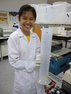
A 2 litre measuring cylinder. HUGE.

RUNNING MAN! Just kidding. XD

The anatomy of a mouse.

??????
We felt great about today. Although it ended later than usual, at least we got to do some hands-on like when we were casting the gel. Other than continuing the western blotting, we will also be going through flowed cytometry. Looking forward to a new day!
No comments:
Post a Comment